|
Case
An eighty five year old female visited to our hospital for evaluation of liver cirrhosis.
Routine ECG(Fig. 1) and simple chest X-ray(Fig. 2) were recorded. She was diagnosed as "situs inversus" finally.
Legend
|
Fig 1A
Conventionally recorded ECG showed abnormal axis deviation of P wave, QRS complex and T wave in limb leads and abnormal R wave progression in precordial leads.
|
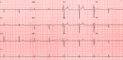 |
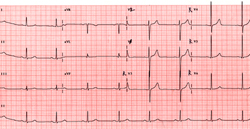
Fig 1B
ECG was recorded in limb leads after both arm leads were reversed and in right sided precordial leads.
|
 |
|
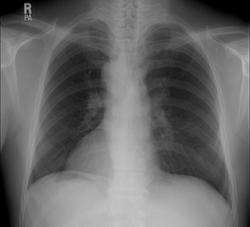
Fig 2.
Simple chest X-ray showed dextrocardia, long hypoarterial right main bronchus, left sided large lobe of liver and right sided stomach gas.
|
|
자료 제공 :
성균관의대 내과학교실 박승우교수
☞ 질문이나 의견이 있으시면 회원들의 공간
 에 글을 써주시기 바랍니다 에 글을 써주시기 바랍니다
|