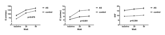
Background: To evaluate temporal changes of left ventricular (LV) geometry and diastolic function after aortic valve replacement (AVR) in patients with aortic stenosis (AS), 38 patients (mean age, 57.8¬±12.1 years) underwent low-level supine bicycle exercise Doppler echocardiography (EDE) 59.2¬±33.0 months after AVR. During EDE, early diastolic peak velocity of transmitral flow (E) and annular movement (EвАЩ) was measured. EDE was performed in 19 sex- and age-matched controls for comparison. After AVR, LV mass index (LVMI)/LV end-diastolic volume index (LVEDVI) decreased up to 27.4¬±32.7%. Although pre-AVR E velocity was similar, pre-AVR EвАЩ velocity was lower (4.3¬±1.6 versus 7.7¬±1.6 cm/sec, p<0.005) which resulted in higher E/EвАЩ in AS patients (16.7¬±5.4 versus 9.3¬±1.8, p<0.001). Pre-AVR LVMI/LVEDVI showed negative correlation with EвАЩ (r=0.499, p<0.001) and positive correlation with E/EвАЩ (r=0.376, p=0.011). E/EвАЩ did not change significantly after AVR (from 16.7¬±5.4 to 15.5¬±4.7, p=0.573) despite EвАЩ increase after AVR (from 4.3¬±1.6 to 6.3¬±1.8 cm/sec, p<0.001). Both E and EвАЩ increased progressively with exercise and the degree of E velocity increase during exercise was not different (p=0.675), whereas EвАЩ velocity increase was more prominent in normal controls than in AS patients (p<0.001). The peak E/EвАЩ during exercise was higher in AS patients (17.8¬±5.0 versus 10.4¬±1.7, p<0.05). Prevalence of patients with peak E/EвАЩ>13 during EDE was higher in AS patients (89.5% [34/39] versus 0% [0/19], p<0.001). When used separately in multivariate analysis, both pre-AVR and post-AVR LVMI/LVEDVI were the only independent factor associated with peak E/EвАЩ during EDE. Conclusions Persistent LV diastolic dysfunction exists up to 5 years after uneventful AVR and failure in normal physiologic augmentation of LV relaxation during exercise associated with incomplete or inadequate regression of LVH is the main mechanism
|