| єя«•«ьљƒ : ∆чљЇ≈Ќ
|
ЅҐЉцєш»£ - 540629 97 |
| Comparison of IVUS Findings Using Different Tissue Characterization System; VH-IVUS vs. iMAP IVUS |
| мЭЄм†ЬмЭШлМА мЭЉмВ∞л∞±л≥СмЫР¬є , м§СмХЩлМАнХЩкµРл≥СмЫР ¬≤ |
| мЭімД±мЬ§¬є, лПДм§АнШХ¬є , кєАмГБмЪ±¬≤ , мµЬнШДлѓЉ¬є , кґМмД±мЪ±¬є , лВ®кґБм§А¬є , к≥љмЮђмІД¬є , мЭімЫРл°Ь¬є |
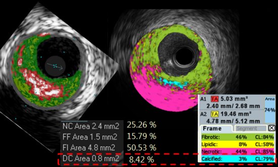
Background: Two different tissue characterization techniques are available in the real world IUVS-guided percutaneous coronary intervention (PCI), VH-IVUS gave clinical implication especially identification of thin-cap fibroatheroma that can predict clinical event. The iMAP IVUS system is relatively new technology available since early this year. Limited data associated with iMAP IVUS and no systemized report for comparison of two different systems.
Methods: We performed pre-PCI IVUS using both VH-IVUS and iMAP IVUS system consecutively for 20 patients with coronary artery disease (13 stable angina, 7 acute coronary syndrome) treated with stent implantation. We selected and compared 40 pairs of cross-sectional tissue characterization IVUS images.
Results: The iMAP-IVUS interpreted as less calcium and necrotic area comparing VH-IVUS in lesions without superficial calcium and acoustic shadow. Nearly all ultrasound acoustic shadows such as superficial calcium, attenuated plaque and even wire artifact were interpreted as necrotic core with high confidence level in iMAP IVUS, whereas VH-IVUS could discriminate tissue type in area behind calcium. VH-IVUS also interpreted wire artifact as necrotic core in PCI using parallel wire technique. VH-IVUS showed heavily necrotic area around stent metal, but no peri-stent metal necrotic core were found with iMAP IVUS. Intraluminal mass (maybe thrombus) was interpreted as fibro-necrotic lesion in iMAP IVUS using the analysis option for lesion of interest, mainly fibrotic in VH-IVUS. Exact concordant level of cross-sectional IVUS image could not compared in few cases because VH-IVUS images were captured by EKG gating whereas iMAP IVUS by 2 frames/mm interval.
Conclusion: There were significant discrepancies between two tissue characterization IVUS systems, especially all lesions with acoustic shadow were interpreted as necrotic core by iMAP IVUS. We need more detailed study such as pathology to verify the accuracy of these two systems.
|
|
|
Warning: getimagesize(/home/virtual/circulationadmin/renewal/econgress/conference/abstract/img_files/aaa.jpg) [function.getimagesize]: failed to open stream: No such file or directory in /home/virtual/circulationadmin/new/econgress/conference/manage/schedule/view_abstract.php on line 164
|
|


