| єя«•«ьљƒ : јюјЇњђ±ЄјЏїу
|
ЅҐЉцєш»£ - 540472 10 |
| Extent of Late Gadolinium Enhancement in Cardiac Magnetic Resonance and Its Relationship with Left Ventricular Longitudinal Functional Reserve During Exercise in Patients with Hypertrophic Cardiomyopathy |
| Cardiology Division, Yonsei University College of Medicine¬є , Department of Radiology, Yonsei University College of Medicine¬≤ |
| лђЄм†ХкЈЉ¬є, мЦСмЪ∞мЭЄ¬є , м°∞мЭЄм†Х¬є , мЛђмІАмШБ¬є , кєАмШБмІД¬≤ , мЮ•мЦСмИШ¬є , м†ХлВ®мЛЭ¬є , м°∞мКємЧ∞¬є , нХШмҐЕмЫР¬є |
BACKGROUND: Late gadolinium enhancement (LGE) is a common abnormal observation in gadolinium-enhanced cardiac magnetic resonance (CMR) in hypertrophic cardiomyopathy (HCM), reflecting myocardial fibrosis. We hypothesized that the extent of LGE would correlate with parameters of left ventricular (LV) functional reserve during exercise in HCM patients.
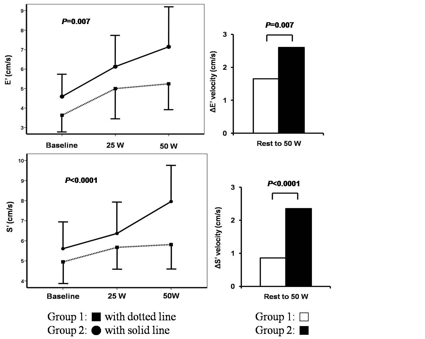
METHODS: Mitral septal annular systolic (SвАЩ) and early diastolic (EвАЩ) velocities were measured at rest and during graded supine bicycle exercise (25W, 3-minute increments) in 46 HCM patients (32 male, mean age 53 years). LV longitudinal diastolic and systolic functional reserve indices were calculated as follows; diastolic function reserve index=ќФEвАЩ√ЧEвАЩbase, where ќФEвАЩ is the change in EвАЩ from baseline to 50 W of exercise, systolic reserve index=ќФSвАЩ√ЧSвАЩbase, where ќФSвАЩ is the change in SвАЩ from baseline to 50 W of exercise. The degree of myocardial fibrosis was assessed with LGE by CMR. The patients were divided into two groups according to the extent of LGE (% of DE mass to LV mass [range; 0 to 37%, the median=6%]): Group 1 (n=23) with larger extent of DE (LV with DE вЙ• 6%) and Group 2 with lesser DE.
RESULTS: There were no significant differences in resting echocardiographic parameters between two groups except SвАЩ (5.0¬±1.0 vs. 5.8¬±1.2 cm/s [P=0.022]) and deceleration time of mitral inflow (230¬±75 vs. 190¬±36 ms [P=0.023]). However, increments of EвАЩ and SвАЩ during exercise were significantly smaller in Group 1 (ќФEвАЩ: 1.5¬±1.0 vs. 2.8¬±1.8 cm/s [P=0.007]; ќФSвАЩ: 0.9¬±0.8 vs. 2.2¬±1.2 cm/s [P<0.0001]). In addition, LV functional reserve indices were significantly lower in Group 1 when compared to Group 2 (diastolic index: 5.5¬±3.4 vs. 12.8¬±7.7 [P=0.001]; systolic index: 4.7¬±4.5 vs. 12.6¬±7.4 [P<0.0001]).
CONCLUSION: Augmentation of LV longitudinal function during exercise is more blunted in HCM patients with greater extent of LGE on CMR. The extent of myocardial fibrosis might be a pathologic substrate which determines LV functional reserve during exercise in HCM.
|
|
|
Warning: getimagesize(/home/virtual/circulationadmin/renewal/econgress/conference/abstract/img_files/AHAfigure.jpg) [function.getimagesize]: failed to open stream: No such file or directory in /home/virtual/circulationadmin/new/econgress/conference/manage/schedule/view_abstract.php on line 164
|
|


