| єя«•«ьљƒ : ±Єњђ
|
ЅҐЉцєш»£ - 530496 337 |
| Local hemodynamic changes caused by main branch stent implantation and subsequent side branch balloon angioplasty in a coronary bifurcation lesion |
| мДЬмЪЄлМАнХЩкµР л≥СмЫР¬є , Marquette Univeristy¬≤, Stanford University Medical Center¬≥ |
| кµђл≥ЄкґМ¬є, Andrew R. Williams¬≤ , John F LaDisa, Jr¬≤ , Peter J Fitzgerald¬≥ , кєАнЪ®мИШ¬є, л∞ХмШБл∞∞¬є |
Backgrounds: Abnormal blood flow patterns promote inflammation, cellular proliferation and thrombosis and can be established by local geometric changes after stent implantation. We sought to quantify altered hemodynamics due to main branch (MB) stenting in a coronary bifurcation and to evaluate whether subsequent side branch (SB) angioplasty can improve hemodynamic profiles.
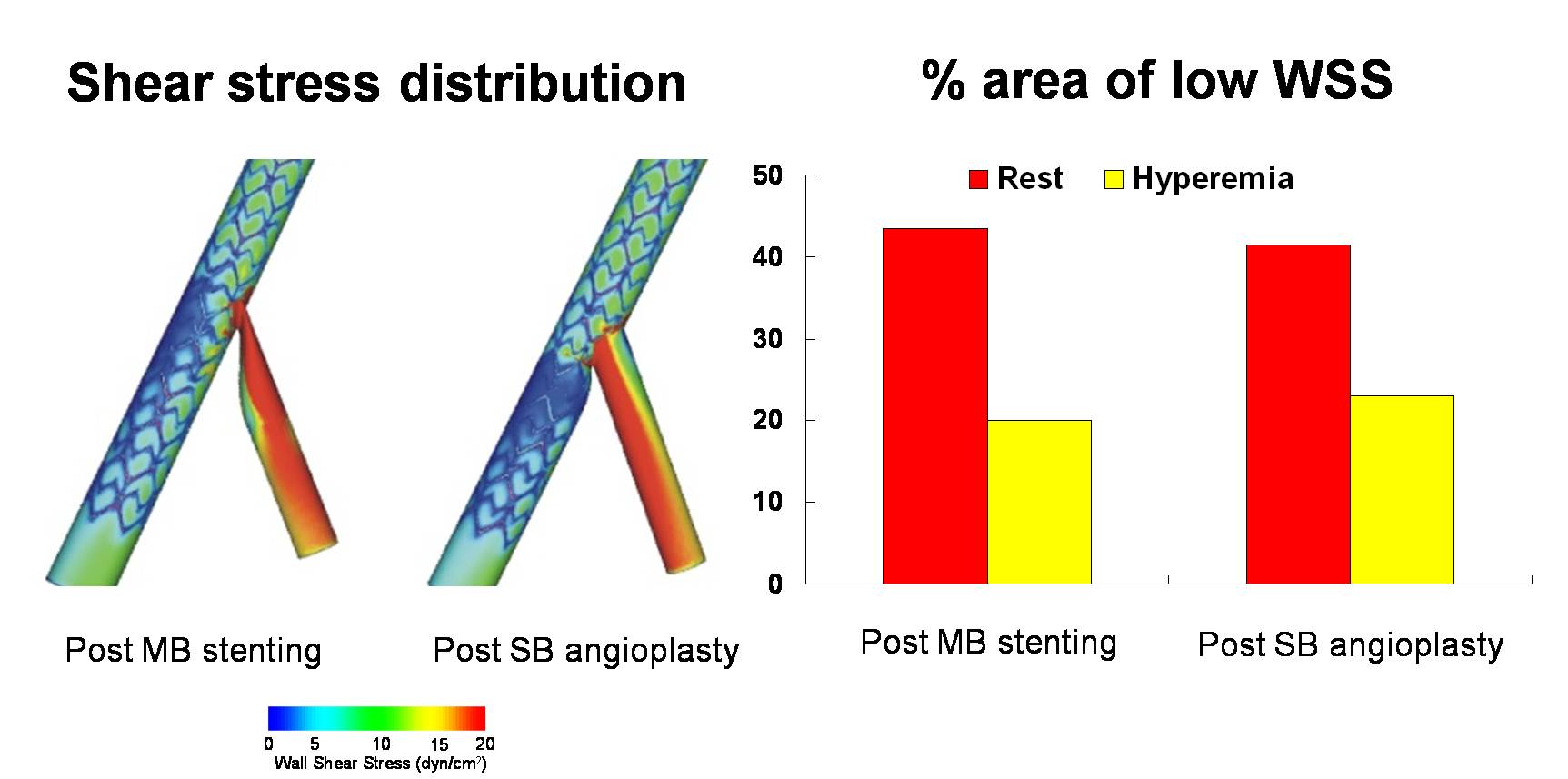
Methods: Computation fluid dynamics (CFD) models were generated from representative intravascular ultrasound images. Fractal ratio of mother and daughter branches was 0.678. Abnormal blood flow patterns were quantified by time-averaged wall shear stress (TAWSS) and oscillatory shear index (OSI) in both resting and hyperemic conditions. TAWSS, OSI and fractional flow reserve were measured before and after MB stent implantatioin and after SB angioplasty.
Results: MB stenting introduced eccentric areas of low TAWSS along the lateral wall of the MB. SB angioplasty resulted in a more concentric region of low TAWSS in the MB distal to the carina and along the lateral wall of the SB. SB fractional flow reserve was >0.9 in all models. MB stenting increased the luminal surface area of low TAWSS (1% to 21%) and this was not improved by SB angioplasty (21% to 23%). Both sites and changes of elevated OSI coincided with those of low TAWSS.
Conclusions: These findings indicate that the most commonly used percutaneous interventional strategy for a bifurcation lesion causes abnormal local hemodynamic conditions. These results may partially explain the high clinical event rates in bifurcation lesions.
Figure. Local flow patterns after main branch stenting and side branch angioplasty (left) and the percentage of the luminal surface area exposed to low time-averaged wall shear stress (< 4 dyn/cm2, right). MB: main branch, SB: side branch
|
|
|
Warning: getimagesize(/home/virtual/circulationadmin/renewal/econgress/conference/abstract/img_files/bkk3.jpg) [function.getimagesize]: failed to open stream: No such file or directory in /home/virtual/circulationadmin/new/econgress/conference/manage/schedule/view_abstract.php on line 164
|
|


