Background: Marfan syndrome (MFS) is a multi-systemic connective tissue disorder associated with mutation of fibrillin-1, the main component of microfibrils. Fibrilin-1 gene mutation could affect the property of carotid arterial wall. We sought to investigate carotid arterial mechanics using velocity vector imaging (VVI) in patients with MFS.
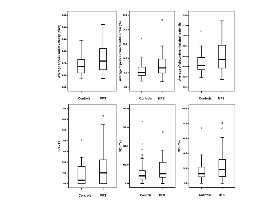
Method: Thirty-eight MFS patients (23 males, mean age 39 ± 10) and 38 age, gender and body surface area-matched controls were studied. Transverse images of both common carotid arteries (CCA) distal to bifurcation were obtained and divided into 6 segments. The peak radial velocity, circumferential strain and strain rate of 6 segments were analyzed using VVI (Figure). The time to peak radial velocity (Ts), circumferential strain (Tst) and stain rate (Tsr) of 6 segments were calculated. Intima-medial thickness (IMT) was also measured.
Results: Although IMT of both CCA was comparable between two groups, average of peak radial velocities, circumferential stain and strain rates were higher in patients with MFS (p=0.004; p=0.059; p=0.029). Moreover, SD of Ts and Tsr were also higher in patients with MFS (p=0.047 and p=0.040) (Figure).
Conclusions: In patients with MFS, carotid artery showed more dynamic, but dyssynchronous arterial expansion during systole when compared with healthy controls. Arterial assessment using VVI may represent a new method for quantifying vascular alteration associated with MFS noninvasively.
|