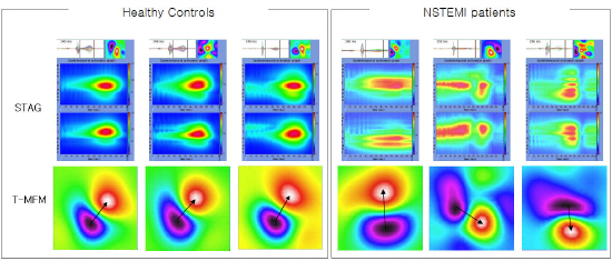
The purpose of this study is to show the difference between healthy controls and severe ischemic patients among unstable angina (UA) and Non-ST-segment elevation myocardial infarction (NSTEMI) patients using the 64 channel magnetocardiography (MCG). We used three methods together to show the difference; 10 MCG parameters, magnetic field map shapes at T-wave peak (T-MFM), and spatiotemporal activation graphs (STAG) during repolarization from J-point to the end of T-wave. MCG data from 4 groups including 185 young controls (YC), 19 age-matched controls (AMC), 110 UA, and 83 NSTEMI were analyzed. As results, 10 parameters showed significant differences (p<0.001) between both controls and NSTEMI patients, and between AMC and UA in six parameters (p<0.05). Among them, after random selection, we compared twenty NSTEMI patients, 15 young and 13 age-matched control subjects (AMC). All controls (100%) showed under 3 MCG parameters that fall out of the normal ranges and, in contrast, 19 NSTEMI (95%) showed over 4 to 10 parameters. STAGs and T-MFM also showed clear difference between controls and NSTEMI showing compressed, prolonged, and/or rotated patterns which should be alternatively created by survived cells after myocardial infarction. These results suggest that changes in the cardiac electrical pathway after myocardial infarction or severe stenosis can be measured using MCG.
|