Background: Left atrial blood flow is optimized to facilitate efficient LV filling during diastole. Vortices in the left atrium (LA) have specific geometry and anatomical location which are critical determinants of directed blood flow during LV filling and ejection. The aim of this study was to characterize left atrial vortex flow pattern using transesophageal contrast echocardiography in normals and patients with atrial fibrillation (AF).
Methods: 25 normal subjects and 35 patients with AF underwent transesophageal contrast echocardiography with slow infusion of Definity and imaged at a mechanical index of 0.4-0.6 in the horizontal chest (0¬Ї), longitudinal chest (90¬Ї) and long cardiac view (135¬Ї). The two components of the velocity vector (angle-independent) on the scan-plane were estimated by a particle image velocimetry (PIV), combined with a Feature Tracking Algorithm. We compared morphology and pulsatility of LA vortex flow between normals and patients with AF.
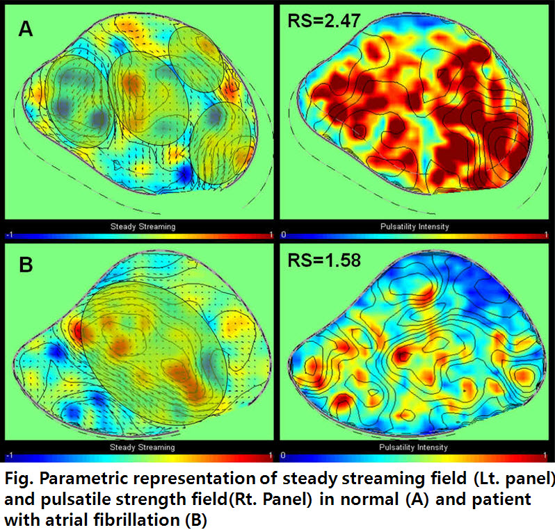
Results: Flow data in freeze frames represent the velocity vector on the scan-plane. In normals, dynamic, compact and multiple elliptical vortices were seen in the entire LA during cardiac cycle. This vortex was persisitent during systole directs vectors towards the left ventricular inflow from pulmonary veins (Fig.A). In patients with AF, echo showd a large, merged, round shaped vortex in the center of LA (Fig.B. Lt. panel). In pulsatility map (Fig. Rt. panel) the relative strength (RS) which represents pulsatility power of LA vortex was significantly lower in patients with AF than normals (RS=1.79±0.33 vs 2.14±0.14, P<0.001, respectively).
Conclusion: It is feasible to characterize and quantify the LA vortex flow using transesophageal contrast echocardiography. It is the principal quantity to recognize the flow structure of LA, and thus offer a new method for early detection of LA dysfunction and potential application for the decision of treatment strategy and guideline of anticoagulation treatment in patients with AF.
|