Background: A previous study suggested that the mechanisms of restenosis in side branch ostium were suboptimal stent expansion and neointimal proliferation. This study expanded the analysis to all the segments after the bifurcation stenting.
Methods: This study examined 73 bifurcation lesions with post-procedural and 9-month follow-up intravascular ultrasound (IVUS) images for both main branch (MB) and side branch (SB). All lesions were treated with drug-eluting stents using the 2-stent T-stenting technique. The IVUS analysis included 5 distinct segments: parent vessel (PV), MB os, distal MB, SB os, and distal SB.
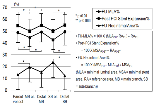
Results: Forty nine lesions (67%) were true bifurcation. Sirolimus-eluting stents were used in 57 lesions (78%). Nine-month angiographic restenosis was noted in 6 lesions (8.2%) in the MB and in 9 lesions (12.3%) in the SB. Minimal luminal area percentage in 9-month follow-up IVUS study (FU-MLA%) was significantly lower in the branchial ostium compared to other segments both in MB and SB. Post-procedural stent expansion percentage (post-PCI stent expansion%)was significantly lower, and neointimal area percentage in follow-up IVUS study (FU-neointimal area%) was significantly higher in the each branchial ostium compared to other branchial segments.
Conclusion: Branchial ostium appears to be associated with smaller MLA compared to other segments in the 9-month follow-up study, mostly due to inadequate stent expansion and higher neointimal proliferation.
|