| єя«•«ьљƒ : ±Єњђ
|
ЅҐЉцєш»£ - 530476 344 |
| Mechanism of side branch jailing in bifurcation lesion: Plaque or Carina shift? |
| мДЬмЪЄлМАнХЩкµР л≥СмЫР¬є, к≥Дл™ЕлМАнХЩкµР л≥СмЫР¬≤, мЧ∞мДЄлМАнХЩкµР л≥СмЫР¬≥, мД±кЈ†кіАлМАнХЩкµР мВЉмД±л≥СмЫРвБі, мХДм£ЉлМАнХЩкµР л≥СмЫР5, мДЬмЪЄмЛЬл¶љ л≥ілЭЉлІ§л≥СмЫР6, мДЬмЪЄлМАнХЩкµР лґДлЛєл≥СмЫР7, мЭШл£Мл≥інЧШк≥µлЛ®мЭЉмВ∞л≥СмЫР8, Stanford University Medical Center9 |
| кµђл≥ЄкґМ¬є, к∞ХнШДмЮђ1, кєАнЪ®мИШ1, лВ®м∞љмЪ±2, нЧИмКєнШЄ2, кєАм§СмД†3, мµЬлПЩнЫИ3, мЮ•мЦСмИШ3, нХЬм£ЉмЪ©4, кґМнШДм≤†4, мЬ§л™ЕнШЄ5, нГБмКєм†Ь5, м†ХмЪ∞мШБ6, м°∞мШБмДЭ7, мµЬлПЩм£Љ7, мШ§мД±мІД8, Peter J Fitzgerald9, William F Fearon9 |
Background: The mechanism of side branch (SB) luminal narrowing after main branch (MB) stent implantation in coronary bifurcation lesions is not completely understood.
Objectives: We sought to investigate the mechanism of geometric changes after MB stent implantation in bifurcation lesions using volumetric intravascular ultrasound (IVUS) analysis.
Methods: Seventy seven patients with de novo bifurcation lesions who underwent provisional SB intervention were prospectively enrolled from 8 Korea, Japan and US centers. MB IVUS was performed before and after MB stent implantation. Both quantitative coronary angiography and IVUS analysis were performed by an independent core laboratory at Stanford University Medical Center.
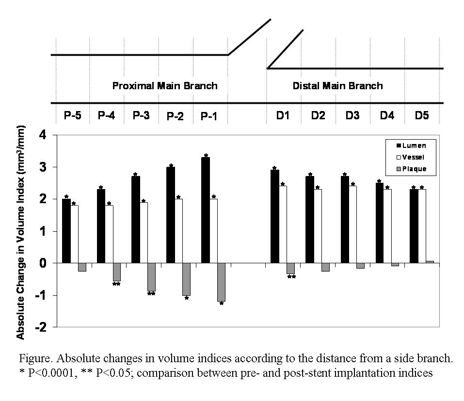
Results: The vessel volume index of both the proximal and distal MB was increased after stent implantation. The plaque volume index decreased in the proximal MB (9.1±3.0 to 8.4±2.4 mm3/mm, p=0.001), implicating plaque shift, but not in the distal MB (5.4±1.8 to 5.3±1.7 mm3/mm, p=0.227), implicating carina shifting to account for the change in vessel size. When each 1-mm volume segment was compared, all changes in vessel and lumen volume indices after stent implantation were significant (p<0.0001). However, the change in plaque volume index was significant only at P-1 to P-4 segments (p<0.0001 in P-1 and P-2, p<0.05 in P-3 and P-4) and D1 segment (p<0.05) (Figure). Plaque volume between segments D2-D5 did not change after stenting.
Conclusions: Both plaque shift from the proximal MB and carina shift contribute to the creation/aggravation of a SB ostial lesion after MB stent implantation.
|
|
|
Warning: getimagesize(/home/virtual/circulationadmin/renewal/econgress/conference/abstract/img_files/bkk1.jpg) [function.getimagesize]: failed to open stream: No such file or directory in /home/virtual/circulationadmin/new/econgress/conference/manage/schedule/view_abstract.php on line 164
|
|


