| єя«•«ьљƒ : ∆чљЇ≈Ќ
|
ЅҐЉцєш»£ - 520890 204 |
| The clinical and angiographic characteristics of coronary perforation during percutaneous intervention |
| лМАкµђк∞АнЖ®л¶≠лМАнХЩл≥СмЫР мИЬнЩШкЄ∞ лВік≥Љ ¬є ,к≥Дл™ЕлМАнХЩкµРлПЩмВ∞мЭШл£МмЫР мИЬнЩШкЄ∞ лВік≥Љ¬≤ ,мШБлВ®лМАнХЩл≥СмЫР мИЬнЩШкЄ∞ лВік≥Љ¬≥ |
| кєАмЖМмЧ∞¬є, кєАкЄ∞мЛЭ¬є ,мЭімШБмИШ¬є ,мЭімІДл∞∞¬є ,л•ШмЮђкЈЉ¬є ,мµЬмІАмЪ©¬є ,мЮ•мД±кµ≠¬є ,м°∞мЬ§к≤љ¬≤ ,лВ®м∞љмЪ±¬≤ ,нЧИмКєнШЄ¬≤ ,мЭімЫРмЮђ¬≥ ,л∞ХмҐЕмД†¬≥ ,кєАмШБм°∞¬≥ |
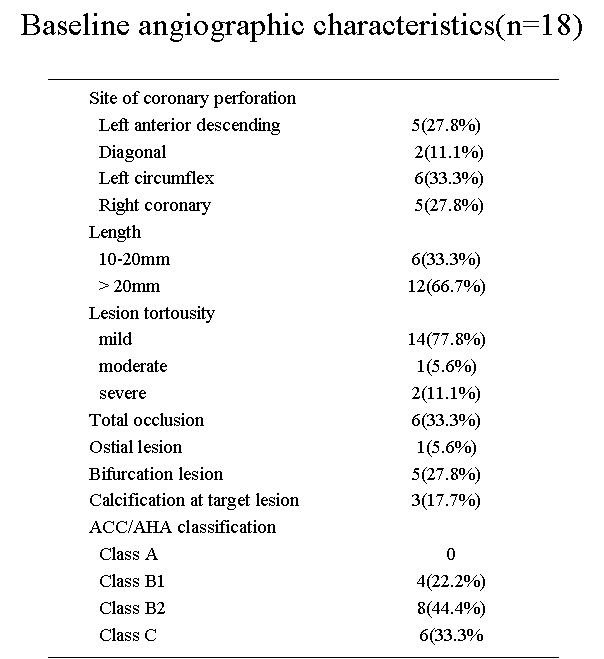
Purpose:Coronary perforation is a rare complication of percutaneous coronary intervention (PCI) with reported incidence from 0.1% to 3.0%. However, it is known to associate with significant morbidity and mortality including cardiac tamponade, emergency coronary artery bypass surgery (CABG), or death. This study is to evaluate the clinical and angiographic characteristics, outcome and management of coronary artery perforation during PCI. Methods:We analyzed patients who had a coronary artery perforation during PCI from 2003 to 2008 in a multicenter. The clinical records, angiographic data were reviewed. Results: Between 2003 and 2008, we experienced 18 cases of coronary perforation in three medical centers. The 11 cases(61%) of all were class III perforation by EllisвАЩs classification. The most common causes of coronary artery perforation was predilational ballooning (50.0%). Most of morphology of lesion was ACC/AHA class B2 or C. Cardiogenic shock or tamponade developed in 6 patients, but nobody was died. Management strategies included medical therapy, ballon, placement of covered or graft stent, and surgery. Conclusions:The coronary perforation might be easily developed in left coronary arteries, longer lesion length and complex lesions more than ACC/AHA class B2.
|
|
|
Warning: getimagesize(/home/virtual/circulationadmin/renewal/econgress/conference/abstract/img_files/perforation.jpg) [function.getimagesize]: failed to open stream: No such file or directory in /home/virtual/circulationadmin/new/econgress/conference/manage/schedule/view_abstract.php on line 164
|
|


