| єя«•«ьљƒ : Clinical award session
|
ЅҐЉцєш»£ - 520019 1 |
| Comparative Analysis between Virtual Histology Intravascular ultrasound and Echoplaque MIB in autopsied coronary arteries: Validation of DICOM-Based Ultrasound Signal Intensity |
| м§СмХЩлМАнХЩкµРл≥СмЫР мИЬнЩШкЄ∞лВік≥Љ мЛђмЮ•мДЉнД∞¬є, Cardiovascular Research Foundation¬≤, Washington Hospital Center¬≥ |
| кєАмГБмЪ±¬є, Gary S. Mintz¬≤, мЭімД±мЬ§¬≥, мЭімЩХмИШ¬є, кєАкЄ∞нЩШ¬є, мДЬкЄ∞мЪ∞¬є, кєАмЭАмШБ¬є, мЭікіСм†Ь¬є, кєАнГЬнШЄ¬є, кєАмєШм†Х¬є, Neil J. Weissman¬≥, л•ШмЩХмД±¬є |
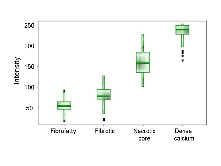
We used histopathologically-validated intravascular Ultrasound (IVUS) Virtual Histology (VH) to evaluate the accuracy of processed reflected grayscale IVUS signal intensity to assess plaque composition. Methods. We harvested 123 coronary arteries from 43 autopsied cases. Greyscale IVUS and VH-IVUS imaging were performed beginning 30mm distal to the ostium of each coronary artery. Greyscale IVUS was processed; and the signal intensity determined from DICOM-stored images using a new MIB system (Echoplaque MIB, Indec, Mountain View, CA, USA). We then compared 436 regions of interest (ROIs). The accuracy rate was expressed using the interpolation method and 95% confidence interval[CI]. Results. Pt age was 49.2±9.12yrs and 82% were males. Four pts had sudden cardiac death and 39 non-cardiac death. The grayscale IVUS signal intensity of VH-IVUS dense calcium was 234.9±19.9 (95% CI: 231~239), of VH-IVUS fibrous plaque was 80.3±20.6 (95% CI: 76~84), and of VH-IVUS fibrofatty plaque was 55.3±14.8 (95% CI: 52~58); however, VH-IVUS necrotic core had a grayscale signal intensity between fibrous and dense calcium - 158.9±31.2 (95% CI:153~165, figure). Correct classification was 89% for dense calcium, 78% for fibrosis, 81% of fibrofatty plaque, and 88% for necrotic core giving an overall accuracy rate of 84%. Conclusion. Plaque characterization using DICOM-based grayscale IVUS signal intensity was comparable with VH-IVUS. This new technique may improve the major limitation of greyscale IVUS, its inability to assess plaque composition.
|
|
|
Warning: getimagesize(/home/virtual/circulationadmin/renewal/econgress/conference/abstract/img_files/mib.jpg) [function.getimagesize]: failed to open stream: No such file or directory in /home/virtual/circulationadmin/new/econgress/conference/manage/schedule/view_abstract.php on line 164
|
|


