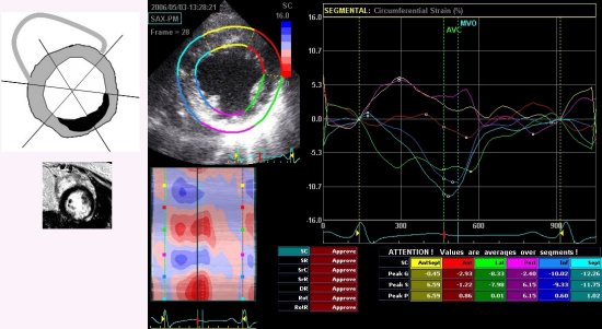
Objectives; The aim of this study was to determine if 2 dimensional strains(2D) correlate with the extent of damaged myocardium measured by contrast-enhanced MRI(ce-MRI) in AMI patients. Background; 2D strains have been introduced as a new noninvasive method to quantitatively assess regional myocardial function without translational motion and tethering effects. Ce-MRI offers high spatial resolution and can identify damaged myocardium using delayed enhancement technique in AMI. Methods; We compared 2D strain patterns with ce-MR images(about within 1 week after onset) in 29 patients with AMI. We evaluated circumferential strain(CS), radial strain(RS) and rotation in short axis view (SAX) using 2D strain analysis of EchoPAC(GE, VIVID 7). In terms of delayed hyperenhancement(DHE) area, we divided 5 groups(group 0, normal : 0%, 1: 0.1-24.9%, 2:25-49.9%, 3:50-74.9%, 4:>75% of DHE). Results; In SAX, The radial strains and circumferential strains were significantly different between groups (p<0.05, ANOVA). However, week correlations was shown only between CS and DHE area(r=0.449, p<0.001). We got the cuff-point value of CS > -17.00 s-1 for damaged myocardium by ROC analysis(AUC=0.745, p<0.001, sensitivity 78.9%, specificity 61%). Conclusions; CS could differentiate the damaged myocardium from normal myocardium. It may allow noninvasive, relatively easy determination of the extent damaged myocardium in AMI patients.
|