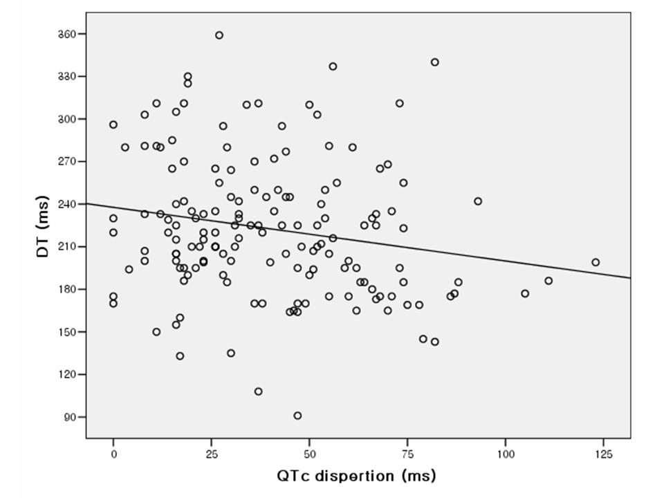
Introduction and Hypothesis: Clinical studies have demonstrated that both QT dispersion and left ventricular filling are linked to myocardial perfusion. We sought to evaluate the relationship between QT dispersion and diastolic parameters in echocardiogram. Methods: QT interval and diastolic parameters were measured in 159 consecutive patients with angina underwent coronary angiography. The patients with regional wall motion abnormlity on echocardiogram were excluded. The measurement of the QT intervals was done automatically by digital QT Guard system (GE Marquette medical system, Milwaukee, USA). The analyzed diastolic parameters of the left ventricle were E velocity (E), A velocity, EA ratio, deceleration time (DT) and isovolumic relaxation time (IVRT). Results: The corrected QT interval (QTc) was correlated with E (r=0.213, P=0.007) and A velocity (r=0.184, P=0.021). The QT dispersion was correlated with E (r=0.249, P=0.002) and DT (r=-0.168, P=0.036). The QTc dispersion was correlated with E (r=0.315, P=0.000), EA ratio (r=0.246, P=0.002) and DT (r=-0.212, P=0.008). Conclusions: Greater dispersion of repolarization is accompanied by changes in the left ventricular diastolic geometry and more restrictive filling. Increased electrical dispersion is related with the left ventricular filling abnormalities.
Figure. Relationship between QTc dispersion and deceleration time
|