| єя«•«ьљƒ : ∆чљЇ≈Ќ
|
ЅҐЉцєш»£ - 490462 49 |
| In Vivo and Ex Vivo X-ray Microscopic Detection of Atherosclerotic Lesions in Apolipoprotein E-Knockout Mice |
| Cardiovascular Center, Korea University, Guro Hospital, Korea1, Institute of Physics, Academia Sinica, Nankang, Taipei, Taiwan2, Department of Materials Science, Pohang University of Science and Technology, Pohang, Korea3 |
| Jin Won Kim1, Hong Seog Seo1, Y. Hwu2, Jung Ho Je3, Chilh Wan Oh1, Seung Woon Rha1, Chang Gyu Park1, Dong Joo Oh1,Young Moo Ro1 |
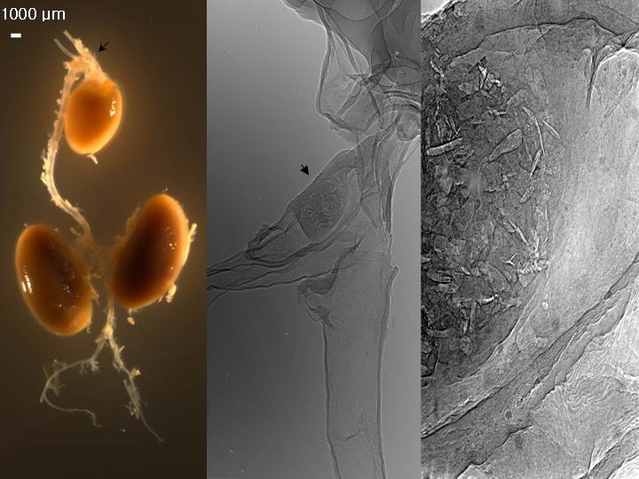
Background: We investigated the feasibility of unmonochromatized synchrotron x-ray to acquire in vivo and ex vivo images of atherosclerotic lesions in apoE-kockout mice. Methods: In the five apoE-knockout mice (apoE-/-, 12, 20, 22, 24, 62-week-old, 3 male) and age/sex matched five wild type mice on cow diets, we acquired in vivo real-time images of thoracic aorta and then, after perfusion-fixation with 10 % formalin, the central arterial trees were dissected intact. Ex vivo synchrotron imagings with tomographic reconstructions were done and compared with pathological findings. Results: For all animals, in vivo real-time images could be acquired without contrast agents despite breathing and heart beating-related movements. In the 62-week-old apoE-knockout mouse, a luminal protruding contour in the ascending aorta was observed and ex vivo images showed the details of atherosclerotic lesions in the ascending aorta without stainings (Figure). The tomographic images of plaque showed central cholesterol clefts as matched with optical images. In the remaining younger mice, in vivo imagings could not detect plaque lesions but ex vivo imagings as well as pathological findings showed the only early atherosclerotic changes as compared with those of control mice. Conclusions: Using unmonochromatized synchrotron radiation, we could detect the advanced atherosclerotic plaque in vivo and depict extremely details of the lesions ex vivo in apoE-knockout mice without contrast agents or stainings.
|
|
|
Warning: getimagesize(/home/virtual/circulationadmin/htdocs/econgress/conference/abstract/img_files/apoEAHA.jpg) [function.getimagesize]: failed to open stream: No such file or directory in /home/virtual/circulationadmin/new/econgress/conference/manage/schedule/view_abstract.php on line 164
|
|


